Critical aspects of ETS

Homepage   

Hyperhidrosis and Facial Blushing


- Hyperreactivity of the Sympathetic Nervous System -


Critical Aspects of Sympathetic Surgery

The confusion surrounding ETS

by Ivo Tarfusser, MD
Updated: 2019-10-20


CONTROVERSIAL ISSUES WITH ETS: INITIAL CONSIDERATIONS  ⇧ 

When searching information about endoscopic thoracic sympathectomy (ETS) or related surgeries on the web or in the scientific literature, it is easy to get confused in the jungle of controversial statements, comments and claims from medical professionals, patients, communities, etc. There has been some effort to standardize certain issues, like the evaluation of quality of life parameters, but there is still too much vagueness when defining details of methods, procedures and outcomes, which makes it almost impossible, not only for patients but also for surgeons, to evaluate and compare methods and results of ETS surgery. This methodological chaos was already mentioned in D. Kopelman and M. Hashmonai's analysis from 2008 ("The Correlation Between the Method of Sympathetic Ablation for Palmar Hyperhidrosis and the Occurrence of Compensatory Hyperhidrosis", World Journal of Surgery, 11/2008, Volume 32, Issue 11).

Judgements on ETS range from miracle cure, with really remarkable results, to anger, regret and condemnation of this surgery. In some countries, the use of ETS has been limited or even prohibited, after a number of patients had suffered or serious complications. For most patients, ETS has represented a significant improvement of their quality of life, but there is a minority of patients who struggle with severe long-term side effects. It is highly likely that this divergence has to be attributed to factors related to the surgical technique, rather than to the patient's individual predisposition for adverse reactions to correctly performed ETS surgery.

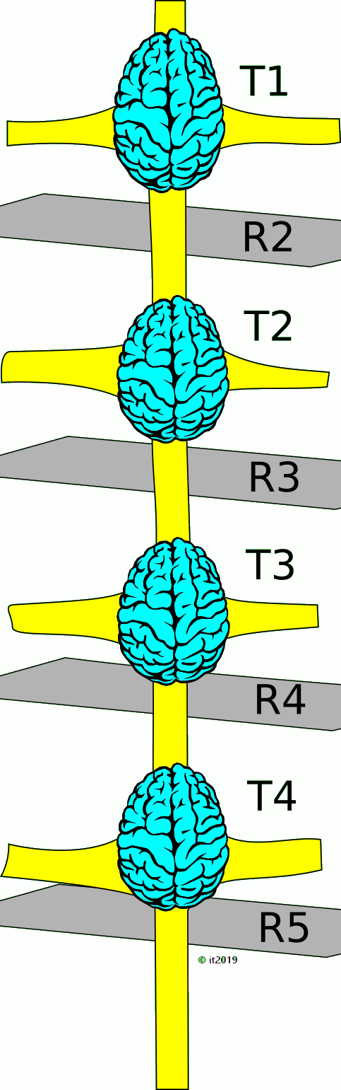
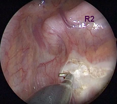
In fact, for the layman (the patient), and not only, it may be astonishing to detect that there are so many different ways to perform ETS surgery. And that there is still an incredible chaos regarding the terminology (e.g. ETS T3, T2, T2+T3, T4, R2, R3, etc.). Even more surprising is that correctly performed procedures (and there are only few of them), observing the principle of minimal trauma at the best level to reach a good result, are reported with the same identical label (e.g. ETS T3) in the literature, as techniques that euphemistically can be described as far from optimum. The latter are with little doubt responsible for many unfavorable outcomes of ETS. Needless to say that this makes it difficult or impossible to evaluate and compare any of the short- and long-term results reported in the majority of the published articles.

Since my fellow colleagues Göran Claes and Christer Drott started pioneering ETS surgery in the late -80ies in Borås, Sweden, the author has been following and performing sympathetic surgery for, carrying out 3500+ ETS procedures in over 1800 patients.

During the first years, the commonly employed technique was a rib-oriented R2-3-4 sympathicotomy (imprecisely called ETS "T2-4"), using blend-mode electrocautery, later revised to R2-3 sympathicotomy (ETS "T2-3").

In the early years, slight to moderate side-effects (especially compensatory sweating) were reported by many patients and considered to be a tollerable and acceptable trade-off to get rid of debilitating focal hyperhidrosis. Severe unfavorable side-effects, particularly compensatory hyperhidrosis and, in rare cases, critical bradycardia, did not become immediately apparent. In the 1990-ies with increasing numbers of surgeries, however, detrimental side-effects turned gradually out to be a potential sequel of ETS in a minority of patients.

As a consequence, the author has ever since focused on analysing critically the technical details of ETS, trying to detect and understand the possible factors responsible for untoward side-effects. Over the years, in my surgical approach, technical changes were sequentially implemented, step by step, in order to correct procedural details that had been identified as potential causes for less favorable outcome. Additional knowledge regarding methodological shortcomings had been gained when re-operating patients, originally operated in other units, in whom the result had not turned out as expected. Modifications that appeared to improve the immediate and, most of all, the long-term clinical result were implemented into the author's surgical routine. Often the improvement was so obvious that a controlled verification study could not be carried out, as it would have required to persevere with methods that had proven inferior or even harmful.

This article, therefore, represents a wrap-up of this personal experience and is meant as a critical overview of available methods. It is not based on orthodox evidence based studies or reviews of published studies. It should try to unravel the incredible confusion that reigns in this particular field of surgery and may contribute to shed some light on why there is such degree of uncertainty and difficulty to compare the results of ETS.


CRUCIAL STEPS AND PITFALLS IN ETS SURGERY  ⇧ 

In most cases, identifying and interrupting the sympathetic chain is a relatively easy task for any thoracic surgeon familiar with thoracoscopic procedures.

Then why is there such a wide-spread variability regarding the incidence of intra-operative complications and the clinical outcome (desired result vs. undesired sequelae)?

It seems reasonable to focus on technical parameters that may explain this issue, since patient related factors should be neutralized by having included significant numbers of patients in the study.

The following factors regarding the interruption of the sympathetic chain may be crucial when analyzing the topic from a methodological point of view:

  1. Where (the exact level)
  2. How (the method used to interrupt the nerve)
  3. What (number of levels neutralized/destroyed)

Among the large number of publications on thoracic sympathectomy, the majority lack an exact description of these elements.

Why is there such a big discrepancy between the outcome in different individuals (besides patient related factors)?

  1. the surgeon makes no effort to respect the boundaries of the ganglia:
    • he focuses on the point where the sympathetic chain crosses the rib to interrupt the nerve
    • In the worst case scenario (and that's not uncommon): he has no knowledge of the location of the ganglia relative to the ribs. It seems incredible, but there are not only a series of peer-reviewed publications, but also published videos, where the author explicitly equates the rib with the location of the ipsinumeric ganglion (e.g. T2=R2, T3=R3 etc.), ignoring the fact that this is never ever the case.
  2. missed level:
    • seems to happen more often than expected (there are at least 2 youtube videos published that show the procedure executed at another level than declared).
  3. the surgeon is unaware or only vaguely aware of the physical properties and biophysical effects of the device he uses to interrupt the nerve
    • He uses a means to interrupt the nerve that causes more extensive damage to the nerve than originally intended.
    • In the worst case scenario (and that's not uncommon): he has no knowledge about the basic physics of electrocautery and, thence, no control over the extension of damage he is causing to the nerve and the adjacent structures

This makes the outcome to a sort of lottery (to the patient): if the anatomical situation has been advantageous, or if mother nature, in the process of healing, has been forgiving, even too radically or less meticulously carried out procedures may yield satisfactory or, at least, acceptable results.

In 2011, the Society of Thoracic Surgeons (STS) published an Expert Consensus for the Surgical Treatment of Hyperhidrosis, attempting to define “best procedures” and to standardize the nomenclature. Unfortunately, the consensus was based on previous publications, most of them reporting the employed surgical technique too vaguely to allow extracting reliable deductions from the published results. Furthermore, in their conclusions, focus has been put on minimizing hypothetical recurrencies, occurring in almost negligible frequency, instead of reducing the impact of side effects (significant incidence). According to this consensus document, the interruption should be rib-oriented and directed to the top of the aimed-at rib. Most astonishingly, ganglion-oriented sympathectomy is considered unreliable due to the variability of the anatomy and location of the ganglia.

This appears to be really remarkable: the sympathetic chain is the target of action and basis of evaluation of the results of surgery, but the recommendation is to aim at and refer to the underlying scheleton without taking care of the anatomy of the nerve; in other words: do not aim at the organ you are intended to operate, because it may shift up and down, aim instead at a nearby structure that seems anatomically easier to identify and more stable in its location (!). Needless to emphasize that this singular concept introduces and consolidates a system error, reproduced by all surgeons that follow these recommendations, which makes it impossible to determine where on the nerve and to what extent the damage has been carried out.

In addition, no critical analysis has been undertaken regarding the different means used to interrupt the nerve and their physical effect on the tissues.

If the objective of the Expert Panel had been to gain a better understanding of the desired and undesired effects of sympathetic surgery, their recommendations will barely reach the goal. The only result would be to universalize the vagueness of the procedures and reports, because based on hitting the sympathetic chain blindly, with whatever method, without knowing which ganglion is severed and to what extent the ganglion is destroyed. The consequence is that most studies will be relatively useless for gaining better knowledge on the subject.

In addition, there is the lack of standard in reporting side-effects, particularly the incidence and the degree of compensatory sweating, spanning over a wide percentage range in different publications.

In the past 20 years, awareness of side effects has become more wide-spread. Attempts have been proposed to reverse the surgery, repair the chain (reconstruction of the sympathetic chain with different techniques), counteract side-effects with drugs and local treatment. However, a detailed critical scrutiny of the surgical techniques has never been endeavoured, to find an explanation for and, eventually, seek to prevent excessively undesirable results.


Anatomical pitfalls  ↰ 

Fig 1: Average location of the ganglia relatively to the ribs. Note particularly the distance between the ganglion and “his” rib, and its close relationship to the next-lower rib.
Fig 2: Higher than average location of the ganglia. Not very frequent pattern of closer to “textbook” anatomy (ganglia mainly confined to intercostal space).
Pic 3: Lower than average location of ganglia. Less common but far from rare pattern with ganglia partly or completely extending over the next-lower rib.

When reading published studies, one get the impression that many surgeons are not aware of the real anatomy of the sympathetic trunk and its variability.

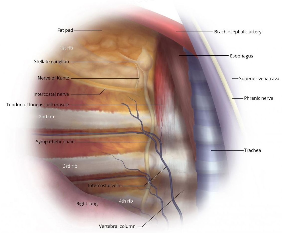
It seems not universally known that a sympathetic ganglion almost always is located in the lower part of the intercostal space, beginning at some distance below the lower edge of the rib carrying the same number, and, more often than not, reaches down to or even covers, partly or completely, the next rib (e.g. the T3 ganglion has no contact with the 3rd rib, but reaches or overlies the 4th rib).

That implies that the widely adopted method to use the ribs as main landmark will interrupt the nerve at a casual level, may fail to target the correct ganglion or sever a ganglion that was not intended to be addressed. The extent of damage to the nerve may be fortuitous and unpredictable, even depending on the technique adopted to interrupt the chain. It may result in partial damage to the ganglion and possible creation of a neurologically unstable ganglion.

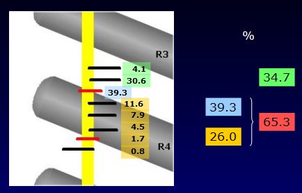
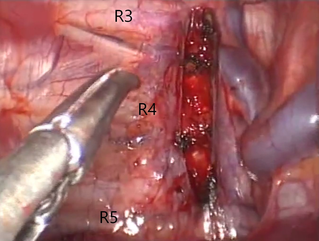
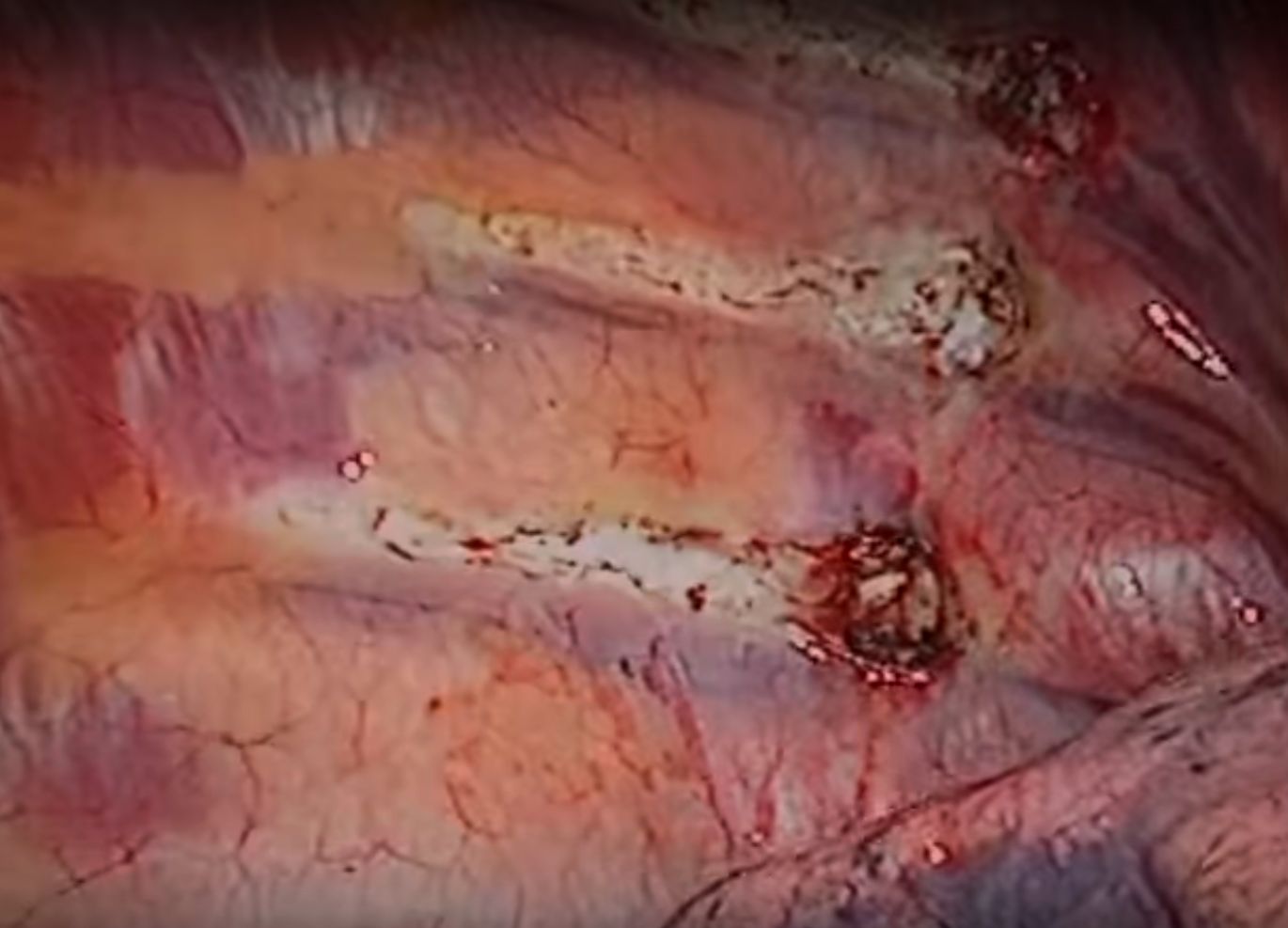
Relationship between the 4th rib and the ganglion T3 at the exit point of the grey ramus communicans, representing roughly the middle of the ganglion (percentage of findings in 121 patients = 242 thoracic cavities) - Study (ISSS Symposium, Vienna 2005). In almost 2/3 of the cases, this connection point is located at or below the 4th rib. In the remainder, around 1/3, the connection was found above the 4th rib, which however does not exclude the lower part of the ganglion reaching or overlapping the rib.

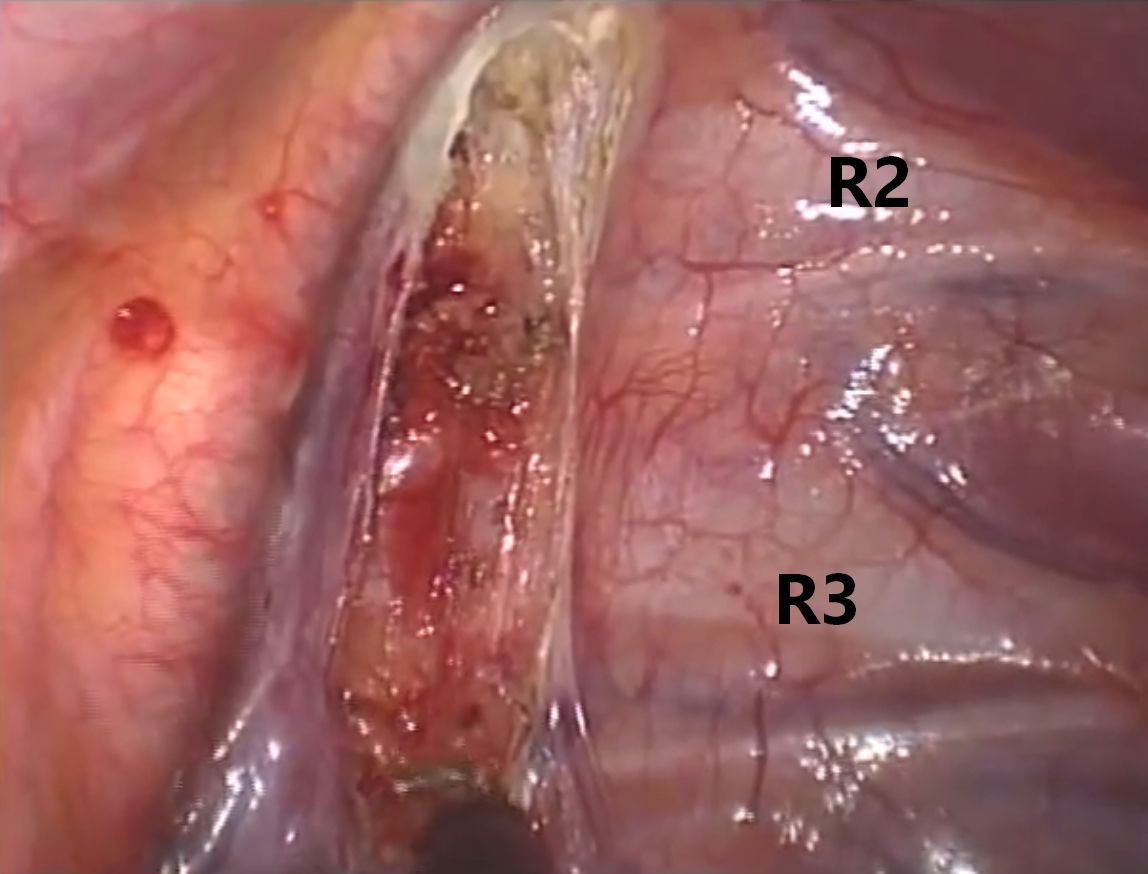
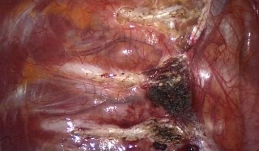
Example of T3 ganglion located on and below the 4th rib.
RC: ramus communicans.
ICN: intercostal nerve
R: rib

Level misjudgment: Another possible pitfall is the wrong interpretation of the level (wrong numbering of ribs), repeatedly documented in published videos (!) in the net. I am afraid that the examples found in these videos are only the tip of the iceberg and, in reality, misinterpretation of the anatomy is far more frequent.

The caption declares division of the chain distal to the T4 ganglion. The video shows clearly that the division was carried out above the 4th rib, resulting in division distal to or through the ganglion T3.
Video


In these two pictures, the 4th rib has been erroneously identified and labeled as the 3rd rib. The uppermost visible rib is clearly the second one. The procedure carried out in this case, visible in the second picture, is a rib-oriented R4 sympathicotomy extended laterally, applying broad cut-coagulation down to the periosteum, apparently chasing hypothetic Kuntz-nerves (an often used, but questionable modus operandi that adds unjustifiable trauma without proven efficacy).
Video

Another example of didactic video on thoracic anatomy with wrong labeling of rib. The rib indicated as the 3rd one is, as a matter of fact, the 4th rib.
Video

Umpteenth example of unbelievable mistake in interpreting the anatomy. The 2nd rib is labeled as 1st rib, and so on downwards. BTW: the video comes from a thoracic surgery unit claiming to have performed thousands of ETS surgeries.

The same website displays the above image that erroneously positions the T1 stellate ganglion above the 1st rib...
In addition, the video shows harsh use of electrocautery (R4 and R5 sympathicotomy) without taking mimimum care about the location and boundaries of the ganglia.
Video

Yet another surgeon exhibiting inadequate knowledge of the anatomy: 2nd rib labeled as 1st rib, 3rd rib labeled as 2nd; grey ramus communicans of T2 ganglion labeled as Kuntz nerve. In addition, the video shows unjustifyable use of fulgurating electrocautery with extensive burn the periosteum of the ribs lateral to the sympathetic chain, seemingly following an almost certainly useless, but unnecessarily traumatic and wide spread habit of chasing hypothetical Kuntz nerve fibers.
Video

The video shows an asymmetric resection, presumably due to misjudgement of levels: resected ganglia T3 and T4 (3rd to 5th rib) on the right; ganglia T2 and T3 on the left (2nd to 4th rib)
Video


Technical pitfalls  ↰ 

A considerable number of surgeons are not familiar with the physics of electrocautery, leading to analogous risks, as mentioned in the preceding paragraph, resulting from the uncritical use of this device:

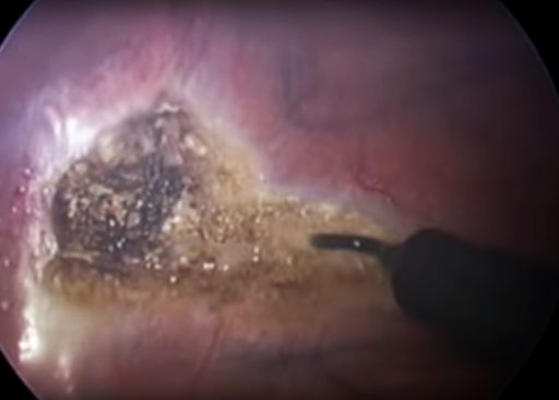
Pure cutting current (continuous unmodulated current >=200V), on the other hand, applied on a minimal surface (needle electrode or tip of L-hook) will cause optimal and concentrated energy delivery to the tissue, resulting in a clean cut with a sealed cut surface and minimal heat penetration into the underlying tissue. At the point of contact, the tissue cells will boil and literally expload in milliseconds, the vapor creates an immediate raise in tissue impedance (resistance) that prevents further penetration of electrical energy and spreading of heat to deeper layers. This electrocautery technique, along with clipping, is more precise, requires relatively low energy and should, therefore, be preferred over other techniques based on heat delivery, including laser.

The same pattern of continuous unmodulated current below 200V will not reach sufficient tissue temperature to vaporize the intracellular water, but leads to Soft Coagulation that spreads gradually through the tissue, without carbonizing it. This technique is the preferred method to stop bleeding in a pinpoint fashion, but should not be used to interrupt the nerve because it is difficult to determine with sufficient precision the extent of thermal damage along the nerve.

Standard Coagulation and Blend mode (cutting + coagulating) requires higher peak voltage settings compared to pure cutting current and has the property to penetrate deeply into the tissue and spread heat uncontrollably along the nerve. Applied long enough, it will carbonize the tissue. Superfluous to mention that its inaccurate use may cause partial thermal damage to the adjacent ganglia or to contiguous structures (intercostal nerve, costovertebral joint etc.), if not employed with extreme caution. Even more dangerous is the use of Fulguration mode in such sensitive environment as the thoracic cavity, characterized by voltage settings as high as to create sparks to reach the tissue even without direct contact. Some surgeons have the (hazardous) habit to apply cut/coag-energy on the electrode while pushing it against the targeted structure (e.g. a nerve), instead of grabbing and lifting it away from the underlying tissue before switching on the current. This technique makes it difficult to limit the depth of mechanical and thermal damage. On the other hand, when pulling the nerve (e.g. with the hook electrode) and applying too low energy, heat will spread rapidly along the nerve and cause more extended necrosis than intended, without or before dividing it.

Frequency: 200 - 3300 kHz (no neurostimulation occurs above 100 kHz)

Nerve tissue is destructed at temperatures at or around 70°C and remains intact at or below 40°C. Unstable nerve tissue due to permanent permeabilization of the axon or cell membrane, an issue not yet addressed in publications dealing with sympathetic surgery, may develop in the layer between necrotic and still healthy tissue, giving rise to chaotic depolarization of nerve fibers and uncontrolled discharge of neurons if the segment includes a ganglion, thus producing pathological nerve signals.


Levels to be targeted  ↰ 

In the early days of ETS (Kux 1977 and later in the 80-ies Claes and Drott), there was a tendency to proceed radically enough in order to guarantee total dryness with least possible risk for recurrence. Consequence of this philosophy was the interruption of the sympathetic chain on different levels, originally severing the sympathetic trunk over the 2nd, 3rd and 4th rib (later in the 90ies reduced to 2nd and 3rd), employing high intensity electrocautery, without identifying the exact location of the ganglia.

Interestingly, this technique is still widely in use, presumably because many surgeons, beginning to perform ETS, rely on the original description of the technique, having in the meanwhile been adopted on thousands of patients.

A turning point has been the report by CC Lin who, besides having introduced the clamping method, postulated the efficacy of the T4 approach for palmar hyperhidrosis, which later was included into the Lin-Telaranta-classification.

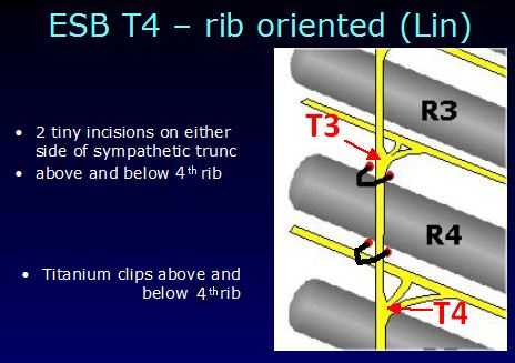
Personally, the author was not able to reproduce the excellent results of ETS T4 reported by Lin. In a series of 16 patients, in whom the T4 ganglion was dissected free, isolated and completely destroyed, the side-effects were admittedly greatly reduced, but the desired effect was not reliable: in some patients, the result was asymmetrical, in some the reduction of sweating was insufficient and others experienced a relaps within months. Over half of the patients required repeat surgery, inducing me to climb a step higher and target the T3 ganglion ever since, focussing exclusively on the ganglion, not on the rib.

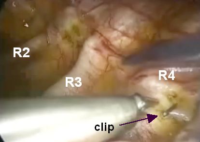
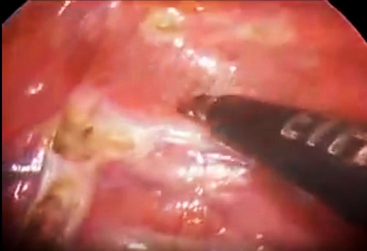
The explanation for this incongruency became clear when observing Lin performing the surgery at the bi-annual symposium on sympathetic surgery in Tampere, Finland, in 2001. He used to place 1 clip above and 1 clip below the 4th rib (Fig.). From an anatomical point of view, the T3 ganglion was compressed by the upper clip, while the T4 ganglion remained completely unharmed by the procedure. This supported the view that isolating and/or destroying the T3 ganglion would be the best compromise to obtain a good and enduring result in patients with hyperhidrosis of the upper extremity, while reducing side-effects to a tollerable minimum. Neutralizing the ganglion was carried out by interrupting the interganglionic segments above and below the ganglion and the grey ramus communicans, using either clean cutting diathermy (with final or careful soft coagulation of the ganglion itself) or by applying clips without destroying the ganglion. No significant difference regarding the result was noted between the two methods.

Surprisingly, apart from improved side effects, the rate of clinically significant recurrence of palmar sweating dropped to almost nil, compared to the T2 method adopted in the preceding decade.

Similarly, the T3 method according to the Lin-Telaranta-classification for facial hyperhidrosis and, especially, facial blushing, did not deliver reliable results when excluding the T3 ganglion alone. Applying the clip or cutting above the 3rd rib may, however, damage the T2 ganglion sufficiently to get the desired effect. Better, though, the interruption carried out above the upper end of the T2 ganglion (corresponding almost always to the lower edge of the 2nd rib) will block all signals directed headwards without damaging the T2 ganglion. However, this will also interrupt a significant number of afferent fibers, deputed to backfeed information about peripheral skin temperature. When the face starts sweating, these fibers report back that skin temperature is beginning to decrease, thus reducing the efferent response to the sweat glands. Disrupting this mechanism may result in more significant reactive sweating, particularly in individuals with an improperly working “thermostat” (like many of those suffering from facial hyperhidrosis at relatively low outside temperatures or after slight physical activity).

Cutting/coagulating/lasering the trunk mid over the anterior surface or on top of the 2nd rib is never required and carries an avoidable risk of damaging the stellate ganglion, particularly in case of a lower than average location of the ganglion (in around 0.3% of cases it covers the anterior face of the rib, partially or completely, resulting inevitably in a Horner's syndrome).


TECHNIQUES IN USE  ⇧ 


A. Interruption of the chain (sympathicotomy and sympathetic block)

Clamping (compression) with clips

Clip correctly placed parallel to pleural surface

 + 
  • no heat damage
  • if limited to single interganglionic segment: potentially reversible
  • low grade trauma
  • short nerve segment engaged
 – 
  • clip can dislocate (especially if applied perpendicular to pleural surface)
    Clip placed perpendicular to pleural surface.

  • aiming at top or mid of rib may sqeeze part of the ganglion and lead to chronic irritating effect on ganglion:
    >> paradoxic reaction
    >> asymmetry

Electrocautery

Plain Cutting Current Mode
Excellent dissection technique: thin hook electrode taking small "bites" of tissue with the tip. Plain cutting energy is concentrated to a tiny surface, leading to immediate vaporization of water at the contact point. Sudden increase of tissue impedance interrupts the current flow within milliseconds and prevents heat propagation to deeper layers. Dissection proceeds in a piecemeal fashion millimeterwise. Note the intact capillaries reaching almost to the cut edge and the minimal rim of coagulation necrosis. No carbonization or signs of thermal damage of underlying structures.
Video
(NB: the video shows a resection of ganglion of T3 and T4, the T2 ganglion is barely touched and not excised, in contrast to what declared in the caption).

 + 
  • very thin coagulated layer at cut edge (thermal seal): prerequisite is the use of a thin-pointed electrode
  • no heat damage on nerve beyond cut edge
  • no heat damage to surrounding tissue and structures
  • if limited to interganglionic segment:
  • low grade trauma
  • only fractions of millimeter of nerve tissue engaged, providing seal
 – 
  • minor bleeding may occur
  • wrong technique or unsuitable electrode: ATTENTION! Broad tissue contact with electrode will cause heat damage extending beyond intended cut line (soft coagulation effect). Too low voltage setting has same effect.
  • aiming cut on rib may cut through ganglion, leaving it partially viable and unstable (similar issue as with clips on ganglion)
Blend Current Mode
Popular among surgeons because of producing cuts with less bleeding due to concomitant coagulation of adjacent tissue. However, the unforeseeable spread of thermal energy makes its use very questionable in sympathetic surgery.
 +  none
 – 
  • extended coagulated layer at cut edge (thermal spread)
  • heat damage along nerve beyond cut edge depending on voltage
  • heat damage to surrounding tissue and structures
Soft Coagulation Mode
Should be used very carefully to stop bleeders, beware of untargeted coagulation
 + 
  • none (for interruption of the nerve)
  • best use for subtile pin-point coagulation of bleeding vessels
 – 
  • will not cut through nerve, but only achieve local nerve tissue necrosis with difficult to control spread of thermal energy along the nerve and to surrounding tissues and structures
Standard Coagulation Mode

Far too often and by too many used to obtain broad coagulation. High settings will have a cutting effect and prolonged energy delivery will carbonize the tissues. This mode should not have any place in surgery on the sympathetic chain, due to its deep and uncontrollably necrotizing effect on nerve and surrounding structures.

The video shows several technical steps that should be avoided: Coagulation current at a far too high setting to obtain cutting effect, in addition very close to the stellate ganglion. Uncritical "cooking" and burning of sympathetic chain and underlying anatomical structures, apparently without caring about the real anatomy and boundaries of the ganglia and the physics of electrocautery.
Video (2009)

Fulguration Mode

Even more dangerous and destructive than standard coagulation. Off limits for ETS.

Wide area fulguration with high voltage setting. Sparks bridge gap to tissue with carbonization and inadequate control over depth effect.
Video

Commonly used when employing ultrathin needle-scope (3 mm) and trocars (2-3 mm) that do not accomodate hook dissectors, but only tiny ball-tip electrodes. One may question the cosmetic advantage of reducing the scar in the skin by 2-3 mm, while risking larger damage inside along the chain...

Laser

Similar thermal effect as blend or fulgurating mode electrocautery with difficult to judge and limit the extent and depth of heat damage.

Cold Cut (e.g. scissors)

 +  delivers clean cut edges with minimal tissue necrosis
 –  lack of seal on cut edge may lead to neuroma formation

Harmonic Scalpel


 +  delivers clean cut edges with minimal tissue necrosis and good seal, similar to plain cutting current
 – 
  • bulky dissecting instrument tips
  • coagulation of bleeders may fail


B. Removal/destruction of part of the chain (sympathectomy)

Principles:
  • Resection (excision)
  • Heat coagulation of a nerve segment after dividing the chain

Excising/destroying part of the sympathetic chain requires extreme attention to detail, in order to avoid an unnecessary long segment and keep traumatization of surrounding and attached structures to a minimum.

The result from resection/destruction of a segment may vary widely depending on

  • the method used to sever the upper and lower end of the resected segment (plain cutting current, blend current mode, high intensity standard coagulation, cold cut, harmonic scalpel)
  • the length of the resected/destroyed segment of the chain
  • the method used to sever the connections (rami communicantes) and isolate the segment (again any of the above methods may be used, with varying risk for causing thermal injury to intercostal nerves, etc.)
  • completeness of ganglion excision/destruction (complete or partial ganglionectomy)

Ganglionectomy of the T3 ganglion (ganglion-oriented, not rib-oriented!) has shown to be effective in close to 100% of patients with palmar or palmo-axillary hyperhidrosis. If damage to the T2 ganglion is avoided, the procedure results in mild or limited side effects and appears to be the best compromise between desired long-term outcome and undesired sequelae.

Any sympathectomy including more than one ganglion is almost never required.

Facial hyperhidrosis and facial blushing should never be treated with resection of any kind.


C. Any combination of the above techniques


LIST OF PROCEDURES  ⇧ 


There is a huge variety of methods that run under the same umbrella term "ETS". The following section lists the most common among the large array of methods employed by different surgeons to carry out ETS. The great number of possible combinations of methods makes it difficult to give an all-comprehensive listing. Only a few of these procedures follow sound neurosurgical and neurophysiological principles (green), others are acceptable but not optimum (yellow). Most procedures cause, however, more damage than necessary and should be avoided, even if they are still widly in use.

RIB-ORIENTED PROCEDURES

A. Interruption (Sympathicotomy or ESB)
  • T2 (rib-oriented = R2)
    • 1 clip on upper margin of 2nd rib
    • 1 clip on lower margin of 2nd rib
    • 2 clips above and below 2nd rib
    • clean cut on upper margin of 2nd rib
    • clean cut on lower margin of 2nd rib
    • clean cut in the middle of 2nd rib
    • soft coagulation in the middle of 2nd rib
    • blend current, standard coagulation or laser in the middle of 2nd rib
  • T3 (rib-oriented = R3)
    • 1 clip on upper margin of 3rd rib
    • 1 clip on lower margin of 3rd rib
    • 2 clips above and below 3rd rib
    • clean cut on top of 3rd rib
    • clean cut on lower margin of 3rd rib
    • clean cut in the middle of 3rd rib
    • soft coagulation in the middle of 3rd rib
    • blend current, standard coagulation or laser on top of 3rd rib
    • blend current, standard coagulation or laser in the middle of 3rd rib
  • T4 (rib-oriented = R4)
    • 1 clip on upper margin of 4th rib
    • 1 clip on lower margin of 4th rib
    • 2 clips above and below 4th rib
    • clean cut on top of 4th rib
    • clean cut on lower margin of 4th rib
    • clean cut in the middle of 4th rib
    • soft coagulation in the middle of 4th rib
    • blend current, standard coagulation or laser on top of 4th rib
    • blend current, standard coagulation or laser in the middle of 4th rib
  • T2-T3 (rib-oriented = R2-R3)
    • combination of any of above methods on 2 levels
  • T2-T4 (rib-oriented = R2-R4)
    • combination of any of above methods on 3 levels
  • T3-T4 (rib-oriented = R3-R4)
    • combination of any of above methods on 2 levels
  • T2-T5 (rib-oriented = R2-R5)
    • combination of any of above methods on 4 levels
  • T3-T5 (rib-oriented = R3-R5)
    • combination of any of above methods on 3 levels
  • T4-T5 (rib-oriented = R4-R5)
    • combination of any of above methods on 2 levels
B. Destruction or Extraction of nerve segment (Sympathectomy)
  • RESECTIONS on any combination of levels
    • Resections after rib-oriented interruption by any combination of above methods

GANGLION-ORIENTED PROCEDURES

Interruption with or without extraction/destruction of nerve segment
  • T2 (ganglion-oriented)
    • 1 interganglionic clip above T2
    • 1 interganglionic clip above and below T2
    • 1 interganglionic clean cut above T2
    • clean cut above and below ganglion T2
    • clean cut above and below ganglion T2 and coagulation of ganglion
    • coagulation of ganglion T2 without cuts
    • resection of ganglion T2
  • T3 (ganglion-oriented)
  • T4 (ganglion-oriented)
    • 1 interganglionic clip above T4
    • 1 interganglionic clip above and below T4
    • 1 interganglionic clean cut above T4
    • clean cut above and below ganglion T4
    • clean cut above and below ganglion T4 and coagulation of ganglion
    • coagulation of ganglion T4 without cuts
    • resection of ganglion T4
  • T2-T3 (ganglion-oriented)
    • combination of any of above methods on 2 levels
  • T3-T4 (ganglion-oriented)
    • combination of any of above methods on 2 levels
  • T2-T4 (ganglion-oriented)
    • combination of any of above methods on 3 levels
  • T3-T5 (ganglion-oriented)
    • combination of any of above methods on 3 levels
  • T2-T5 (ganglion-oriented)
    • combination of any of above methods on 4 levels

PROCEDURES IN DETAIL  ⇧ 

RIB-ORIENTED PROCEDURES  ↰ 

ETS "T2" (R2)
ETS "T3" (R3)
ETS "T4" (R4)
ETS "T2-3" (R2-3)
ETS "T2-4" (R2-4)
ETS "T3-4" (R3-4)
ETS "T2-5" (R2-5), "T3-5" (R3-5), "T4-5" (R4-5)

ETS "T2" (rib-oriented = R2) ↰ 

The surgeon uses the 2nd rib as landmark to interrupt the nerve. The rib is vertically crossed by the nerve fibers that connect the T1 (stellate ganglion) with the T2 ganglion. The position of the T1 ganglion may vary and could be very close to or, exceptionally, even overly the rib, exposing it to risk for damage (Horner's syndrome).

    Clips
  • clip on upper margin of 2nd rib
    Risk for damaging lower portion of T1 (stellate ganglion).
  • clip on lower margin of 2nd rib
    In ganglion-oriented jargon it corresponds to an interganglionic ESB T1-T2; preferred procedure to treat Facial Blushing and Facial Hyperhidrosis.
  • 2 clips above and below 2nd rib
    The upper clip is useless and risks damaging ganglion T1
    Clip at top and bottom edge of 2nd rib + fulguration of pleura and periosteum lateral to the chain (rationale of such traumatization?). Clips applied in an unstable way perpendicular to rib and pointing at lung. Furthermore, as declared in the video, this questionable procedure is performed for treatment of palmar hyperhidrosis (!).
    Video
    Another example: definitively too close on T1, a totally unnecessary risk taking.
    Video
  • Cut (clean cutting current or harmonic scalpel)
  • cut on upper margin of 2nd rib
    Risk for damaging lower portion of T1 (stellate ganglion).
  • cut mid on 2nd rib
    Interganglionic sympathicotomy T1-T2 with slightly higher risk for damaging lower portion of T1 (stellate ganglion) in case of low location of T1.
  • cut on lower margin of 2nd rib
    Interganglionic sympathicotomy T1-T2; procedure to treat Facial Blushing and Facial Hyperhidrosis (clip application may be preferable).
  • Soft coagulation
  • coagulation in the middle of 2nd rib
    The interganglionic segment (T1-2 ) i exsiccated. If the electrode is activated longer than necessary, the heat creeps along the nerve with a certain risk for damaging the lower portion of the stellate ganglion and for creating partial injury to ganglion T2. If the stellate ganglion stretches down to the 2nd rib or even covers its front face (incidence <0.3%), a Horner's syndrome is pre-programmed.
  • Blend current, standard coagulation, fulguration or laser
  • coagulation over the 2nd rib
    This has been a standard procedure in the past, using blend current mode or coagulation at high voltage settings. In the beginning, it was often combined with cut-coag on 3rd rib (see R2-3).
    As a result of this procedure, the interganglionic segment (T1-2) is divided. There is an increased risk for damaging the stellate ganglion (T1) and for causing marginal injury to T2, creating a potentially unstable ganglion. The higher the voltage and the higher the level on the rib, the higher the risk for such destructive effect with complications (Horner's syndrome, heat damage to adjacent structures) and adverse side-effects as possible consequence.
    Massive fulguration on and around the head of the 2nd rib, extended laterally to encompass hypothetic Kuntz' nerves.
    Coagulation at and above the top of the rib implies a high and totally unneeded risk for irritation or damage to the stellate ganglion (T1), potentially leading to and representing the main cause for Horner's syndrome as complication after ETS. Equally useless the lateral extension of thermal trauma.

    Blend cut mode on 2nd rib and heavy coagulation of cut edges, by the author of the video erroneously declared as ”Dividing the T3 level”.
    Video

    Laser fulguration of sympathetic trunk over 2nd rib.
    Uncontrollable coagulation depth due to antegrade energy delivery. Considerable risk for heat spreading along nerve potentially causing thermal damage to stellate ganglion (Horner's syndrome).

ETS "T3" (rib-oriented = R3) ↰ 

In the literature, the "T3 procedure" has become the most frequently mentioned method to treat palmar hyperhidrosis in the past 15 years. Most of the described methods refer to the 3rd rib and do not even come close to the T3 ganglion. The surgeon uses the 3rd rib as landmark to interrupt the nerve. The rib is vertically crossed by the nerve fibers that connect the T2 with the T3 ganglion. The lower segment of the T2 ganglion lies often very close to or covers partially the rib.

    Clips
  • clip on upper margin of 3rd rib
    Risk for squeezing part of T2.
  • clip on lower margin of 3rd rib

    This will block the chain between ganglion T2 and T3 (interganglionic ESB).

    Method used by some for:
     - palmar hyperhidrosis (recurrence rate reported at around 4%)
     - facial hyperhidrosis (insufficient or asymmetric effect in some cases, recurrence possible)
  • 2 clips above and below 3rd rib
    The upper clip is useless and risks damaging ganglion T2.
  • Cut (clean cutting current or harmonic scalpel)
  • cut on upper margin of 3rd rib
    Risk for damaging part of T2.
  • cut on lower margin of 3rd rib
    Interganglionic sympathicotomy T2-T3; by some used for palmar hyperhidrosis (recurrence rate reported at around 4%).

  • cut mid over 3rd rib
    Interganglionic sympathicotomy T2-T3 with slight risk for damage to ganglion T2.
  • cut on upper and lower edge of 3rd rib
    Upper cut makes no sense, but may cause partial damage to ganglion T2.
  • Soft coagulation
  • coagulation in the middle of 3rd rib
    Risk for extended or incomplete heat damage to T2.
  • Blend mode, standard coagulation, fulguration mode, Laser
  • coagulation on top of 3rd rib
    Severe damage to T2 ganglion without guarantee for neutralizing it, instability possible. T3 ganglion remains unharmed.
  • coagulation in the middle of 3rd rib
    Risk to harm T2 ganglion, leaving an unstable focus. T3 ganglion mostly intact.
    Interruption of sympathetic chain on 3rd rib using fulgurating coagulating current, resulting in a wide crater and relevant damage to ganglion T2, whereas ganglion T3 remains presumably (almost) unharmed. Questionable extension of heat damage laterally to include hypothetical Kuntz nerves with their even more hypothetical role in hypothetical relapses of hyperhidrosis. Too much energy applied to divide nerve leads to extensive spread of heat along the nerve ( Video )

    Exaggerated use of fulgurating coagulation over the 3rd rib, resulting in deep carbonization of tissues down to the costovertebral joint and causing a necrotic surface larger than 15x15mm (to interrupt a 2-3 mm thick nerve), extending laterally for another 10 mm.

    Wide-area destruction over 3rd rib by laser
    Video

  • Oddities
  • Combined use of harmonic scalpel and clips
  • The video shows skeletonization of the chain from 3rd to 5th rib using harmonic scalpel, followed by application of numerous clips along the chain, starting in front of the 3rd rib down to squeezing the upper part of the T4 ganglion in the 4th intercostal space. Difficult to interpret the logic (if any) of such overabundance.
    Video

ETS "T4" (rib-oriented = R4) ↰ 

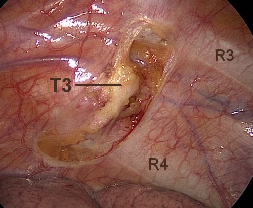
The surgeon uses the 4th rib as landmark to interrupt the nerve. The T3 ganglion in most cases touches the top margin of the rib or covers the rib partly or totally. The T4 ganglion has no contact with the 4th rib: its upper end begins at some distance from the 4th rib's lower margin.

    Clips
  • clip on upper margin of 4th rib
    Partial compression of T3: reasonable effectiveness in palmar hyperhidrosis, but risk for unstable ganglion.
  • Ganglion T3 squeezed by double clips applied above top of 4th rib
    Video

  • clip on lower margin of 4th rib
    Interganglionic ESB T3-T4: uncertain results as treatment for palmar hyperhidrosis (asymmetrical result, significant recurrence rate).
  • Clips above and below 4th rib
    No real rationale when adopted for palmar hyperhidrosis. In this particular case, the clips have been applied after the nerve had been exposed to electrocautery heat and rough mechanical compression. The T3 ganglion ends up heavily crushed, though not completely neutralized. T4 remains unharmed.
  • Cut (clean cutting current or harmonic scalpel)
  • cut on upper margin of 4th rib
    Cut will result close to ganglion T3 or, given the frequent overlap over the rib, more often straight through T3: effective for palmar hyperhidrosis, but risk for unstable ganglion and higher recurrence rate.
  • cut in the middle of 4th rib
    Vague partial damage to ganglion T3 (rationale?).
  • cut on lower margin of 4th rib
    Interganglionic ETS T3-T4: uncertain results regarding palmar hyperhidrosis (asymmetrical result, significant recurrence rate).
  • Plain cut mode, if applied correctly with short impulses on small pieces of tissue at a time, delivers a clean cut and prevents heat from deeper into the tissues and along the nerve. Note the thin coagulated margin of the pleural window and intact capillaries close to the cut edge of the chain.

    Soft coagulation
  • coagulation in the middle of 4th rib
    Partial/extended heat damage to T3 (risk for unstable ganglion), palmar hyperhidrosis reduced to variable extent.
  • Standard coagulation and fulguration mode or Laser
  • coagulation on top of 4th rib
    Severe damage to T3 ganglion without guarantee for neutralizing it, instability not excluded. No impact on T4 ganglion. Palmar hyperhidrosis may be cured, variable degree of compensatory sweating.
  • coagulation in the middle of 4th rib
    Partial/extended heat damage of ganglion T3, in addition even ganglion T4 may be damaged in its upper portion (potentially unstable ganglia).
    Radical coagulation/fulguration of chain over 4th rib, destroying a significant part of the T3 ganglion [rcg3 = grey ramus communicans T3]. The image shows the first step in a more extended procedure (R4-R5 sympathectomy -> Video

ETS "T2-T3" (rib-oriented = R2-R3) ↰ 

Usually performed by severing the sympathetic trunk with electrocautery where it crosses the 2nd and 3rd rib (more or less extended on anterior face of rib).

The cut/coag sympathicotomy R2-R3 (generally, but misleadingly termed ETS T2-T3) was a common surgical method in the 1990-ies and is still widely in use, despite having (or should having) been superseeded by more recent surgical technique.

The potential risks regarding the stellate ganglion during interruption at the 2nd rib level are mentioned above (ETS T2). The procedure leaves the T2 ganglion more or less harmed. Complete neutralization of the T2 ganglion is only guaranteed if the intervening segment stretching over the intercostal space is additionally coagulated or resected. Otherwise, the injury to the T2 ganglion may result partial, possibly leading to a potential hot spot due to chaotic depolarization of unstable nerve tissue membranes. The T3 ganglion is rarely involved (only when using massive coagulation energy over the 3rd rib, the distally located T3 ganglion may suffer marginal damage).

From a therapeutical point of view, there is no rationale today to persevere with this approach. On the contrary, it only may contribute to increase the risk for complications and side-effects.

Bilateral R2-3 sympathicotomy with partial coagulation of ganglion T2. Very audacious or, better, foolhardy use of fulgurating electrocautery on forceps, showing too wide destruction on too many levels. The broad tip requires particularly high voltage settings to obtain the spark jump. Besides the inability to limit range and penetration of thermal energy, causing deeper than required tissue damage with this electrocautery mode, the relatively large uninsulated part of this instrument carries an increased risk for accidental contact and damage to adjacent structures. And, last not least, observing the intrepid grasp and heating of the rostral cut edge of the nerve at the upper margin of the 2nd rib (the video shows it bilaterally !), close to or on the lower part of the T1 ganglion, it may be a miracle if this patient did not end up with a Horner's syndrome. A technique from 1990ies apparently still in use in 2013 (!).
Video (2013)

Bilateral R2-3 sympathicotomy with coagulation of ganglion T2. Another scary video, published in 2018 along with a peer-reviewed article in a pub-med listed journal, showing careless use of electrocautery, apparently due to inadequate insight on the physics of electroinstrumentation and its effect on tissues. Abundant use of fulguration, coagulation while lifting the nerve with uncontrolled diffusion of thermal energy along the nerve, action close to the 2nd rib's upper margin, fierce burning over the rib with the intent to sever any hypothetic Kuntz nerves, in order to prevent even more hypothetic relapses. We hoped that this kind of approach has been superseeded almost 2 decades ago. Long term experience with 8-20% of patients regretting the surgery should have learned us (surgeons) to be more careful when dealing with the sympathetic chain, and maybe prefer the minimal risk of later partial recurrence. Why expose the patient to the risk of thermal injury to the stellate ganglion and a Horner, heat damage on intercostal nerves leading to prolonged neuralgic pain, interruption of the cardiac nerves (bradycardia), partial "boiling" of sympathetic ganglia leaving them crippled and with unforeseeable neurophysiologic reactions. In addition: calling this procedure a T2-T4 approach testifies again the confusion regarding the nomenclature in sympathetic surgery. It appears to be, more correctly, a R2-R3 procedure. The video gives no evidence of the T3 ganglion being even touched.
Video (2018)

This picture shows a curious procedure, involving coagulation of the T2 ganglion from the lower edge of R2 to the middle of R3 and, thereafter, application of clips on both ends (the upper one squeezing the T2 ganglion). The result is a complete exclusion of T2 and no harm to T3.
Video

ETS "T2-T4" (rib-oriented = R2-R4) ↰ 

This triple division of the sympathetic trunk is generally also performed using electrocautery, where the chain crosses the 2nd, 3rd and 4th rib, using varying degrees of thermal energy on the ventral surface of the ribs.

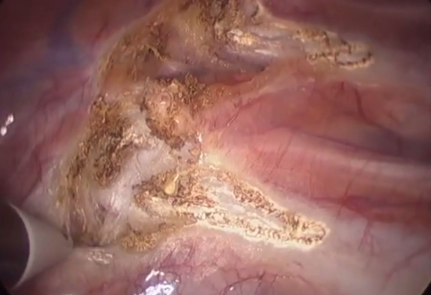
The result is usually partial injury to the T2 and heavy injury to the T3 ganglion, unless the intercostal segments of the sympathetic chain, carrying the two ganglia, are additionally coagulated or in toto excised.

The cut/coag sympathicotomy R2-R4 (generally, but misleadingly termed ETS T2-T4) was the most common surgical method in the late 1980-ies and at the beginning of the 1990-ies. There is evidence (e.g. Youtube videos) that it is still in use in a series of thoracic surgery units around the globe, presumably because of many surgeons basing their procedures on publications dating back to the initial pioneering era.

The risks are the sum of risks mentioned above for the single level rib-oriented procedures.

Today, there is no indicaton for exposing the patient to such traumatic practice and should be definitively abandoned.

Blend-cut and coagulation on R2-R4: early method (late 1980ies, 1990ies)
The procedure shown in the video is performed using a grab-and-burn-technique with a coarse grasper forceps. The ganglia T2 and T3 are both partly damaged, but none has been completely neutralized. The contact surface with the nerve is far too broad, requiring high energy to obtain the division of the nerve. In addition, the long segment without insulation increases the risk for accidental injury to contiguous structures.
Video

Similar blend-cut on rib 2-4. Fulguration extended laterally on rib (questionable practice to divide hypothetic Kuntz nerves)
Video

Curious overenthusiastic „superclipping“ from 2nd to 4th rib: after very rough dissection, 9 clips are applied,seemingly haphazardly, along the intervening sympathetic trunk.
Not only the interganglionic segments, but also the ganglia themselves are squeezed. One may question the rationale of using clips if the ganglia are irrecoverably damaged.
Furthermore, the clips are set perpendiculary to the pleural surface, thereby risking to pinch or cause irritation to the underlying intercostal nerve.
The lateral extension of electrocautery on the ribs ("Anti-Kuntz-Nerve-Extension") adds to the trauma and to postoperative discomfort, but barely to the clinical result.
Video

Mixed procedure with harmonic scalpel: sympathicotomy over necks of 2nd to 4th rib; exstirpation of segment over 4th rib, incompletely injuring ganglion T3. Partly damaged ganglion T2. Main trunk over R2 not hit (deliberately avoided or missed?), just some timid coagulation attempts laterally on 2nd rib (Kuntz hunting?). Logic behind this procedure remains unclear.
Video

An example of how the procedure can be overdone, employing a da Vinci system for robotic assisted surgery that can be performed with much less complex and even more precise thoracoscopic technique. Further remarks:
 1. Resecting or damaging 3 ganglia to treat palmar hyperhidrosis has been abandoned many years ago by surgeons with long time ETS experience.
 2. In particular, the T2 ganglion should not have any place in the primary surgical treatment of palmar hyperhidrosis. Lifting the whole nerve with the hook while applying high voltage diathermy to divide the nerve close to the upper margin of the 2nd rib may injure the T1 (stellate) ganglion, maybe leading to a Horner's syndrome, especially if it is located close to or on the 2nd rib.
 3. The video shows use of too high levels of electrocautery energy with risk for damage to the 2nd intercostal nerve.
Typically, underestimating the incidence of debilitating compensatory hyperhidrosis after too radically or too sloppily performed ETS, in order to prevent the much lower risk for recurrence, is the most frequent mistake of surgeons with limited experience with ETS, inadequate understanding of the anatomy and neurophysiology of the sympathetic chain and/or lack of knowledge about the thermophysical laws and of electrocautery and its effect on tissues.
Video

Horrendous example of extensive carbonizing coagulation on three levels (2nd, 3rd and 4th rib). The action is extended 2-3 cm laterally, apparently to include hypothetical Kuntz nerves. Superfluous to say that the ganglia may have suffered different degrees of damage without being definitively neutralized. Surrounding structures (costovertebral joints, intercostal nerves,...) are needlessly injured. After surgery, the patient suffered severe intercostal and respiratory pain for weeks and developed debilitating compensatory hyperhidrosis.
Video

ETS "T3-T4" (rib-oriented = R3-R4) ↰ 

Generally performed by severing the sympathetic trunk with electrocautery where it crosses the 3rd and 4th rib, delivering varying levels of thermal energy to the anterior face of the ribs.

The result on the sympathetic trunk is a partial, subtotal or complete destruction of ganglion T3, due to proceeding on the 4th (!) rib, since the T3 ganglion often extends down to or over the 4th rib. Severing the nerve at the 3rd rib level will, in the best of cases, divide the interganglionic segment T2-T3; more often, the anatomical situation is less favorable and the T2 ganglion could suffer damage.

Video 
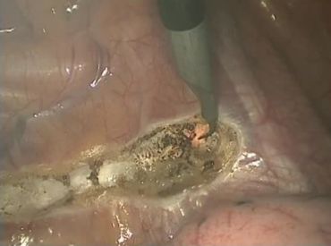
showing bilateral sympathicotomy on both R3 and R4 levels with harmonic scalpel. Since ganglion boundaries are not respected, ganglion T3 is partially injured (maybe also T2).

Excising the intervening segment will generally encompass the T3 ganglion. Exceptionally, in case of low-sitting T3 ganglion, only the upper portion of the ganglion will be involved. The T4 ganglion remains practically always untouched. Exact location and mode of division (scissors or type/energy of current), as well as coagulation of bleeding vessels determine possible extended damage to neighboring ganglia (T2, T4) and thermal injury to intercostal nerves.

As any rib-oriented procedure, the target (i.e. the sympathetic chain) is hit fortuitously, disregarding the anatomical boundaries of ganglia and interganglionic segments.


ETS "T2-T5" (rib-oriented = R2-R5), "T3-T5" (= R3-R5) and "T4-T5" (= R4-R5) ↰ 

Including the R5 level is supposed to increase the chance to treat axillary hyperhidrosis.

Anatomically, it would cause variable damage to the T4 ganglion (without involving the T5 ganglion).

Controlled evidence is lacking that would prove that resecting or coagulating the T4 ganglion only, or in addtion to the T3 ganglion, would be more effective to suppress axillary hyperhidrosis than neutralizing the T3 ganglion alone.

Experience has shown that excluding the T3 ganglion will treat not only palmar, but also coexisting axillary hyperhidrosis with good and enduring effect in most cases.

As always when applying heat energy on - and particularly on top of - the 3rd rib, the T2 ganglion is in likely to be harmed, possibly leading to serious side effects.


Clamping R3-R5. 3 clips applied at top, mid and bottom of rib (eventually from 3rd to 5th rib), after incising the pleura on either side of the chain where it crosses the rib, using blend current with considerable coagulating effect on the nerve. By the way: the professor in his explanations also claims that the ganglia are located on the ribs, i.e. where the nerve crosses and passes in front of the rib. This does not correspond to the real location of the ganglia, which are generally located much lower in the intercostal space. Therefore, clips placed e.g. on the upper and lower margin of the third rib have no effect on the T3 ganglion (which is located much lower, closer to the 4th rib), but are likely to compress part of the T2 ganglion, at least with the clip applied at the upper margin of the rib.
Video

Unusual multilevel clamping R4-6. Clips applied on top of 4th to 6th rib (video shows ganglion T3 overlapping 4th rib and being compressed by 2 clips). Declared as intended for axillary hyperhidrosis, the involvment of the T3 ganglion will however extend the effect also to the hands
Video

R4-R5-Sympathectomy showing wide tissue destruction over ribs and intercostal space, involving the T3 and T4 ganglion as well as intercostal nerves, costovertebral joints, etc., by exaggerated use of electrocautery.
Video


GANGLION-ORIENTED PROCEDURES  ↰ 

ETS T2
ETS T3
ETS T2-3
ETS T3-4
ETS T4

ETS T2 (ganglion-oriented) ↰ 

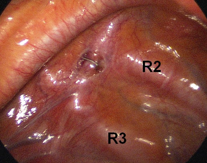
There are various methods to block, destroy or excise the T2 ganglion, but damage or exclusion of the T2 ganglion is almost never required and shoud be avoided. A minority of patients may suffer potentially severe thermoregulatory or cardiovascular side-effects (see conclusions). Only the interganglionic segment between T1 and T2 should be interrupted, taking care to leave both ganglia unharmed. To keep a safe distance to both ganglia, the interruption would have to be executed immediately underneath the lower edge of the 2nd rib. Should reconstruction in case of unbearable side-effects ever be required and attempted, a chance of recovery of the nerve function would only be possible if the interruption had been carried out in this precise location and without damage to the ganglia.

Facial blushing requires blocking the rostrally directed nervous signals exiting T2, which can best be achieved by interrupting the interganglionic segment between T2 and T1 (stellate ganglion), keeping a safe distance to T1. As mentioned, the interruption is best carried out at the lower edge of the 2nd rib, carrying out one single clean cut or, better, applying one single clip).

There is no rationale, whatsoever, to damage the T2 ganglion. Relapses of facial blushing are relatively frequent (in the order of 10%), apparently caused by re-direction of the nervous signals through the stellate ganglion. Missed Kuntz nerves seem to be only a hypothetical cause for recurrencies. The author has performed dozens of re-operations (cut after previous clamping), extending the cut laterally to divide possible Kuntz-nerves, without achieving any improvement in any of the patients. The only procedure apt to resolve the FB in these cases was clamping of the preganglionic branch of T1, but the operation, performed in only 13 cases, carries a significant risk for complications like Horner's syndrome, increased bradycardia, severe CS.

The same procedure will reliably control facial hyperhidrosis, but with a higher risk for postoperative compensatory hyperhidrosis. Patients with this type of HH are often highly thermo-sensitive, their central thermostat being “programmed” to react more intensely to increasing outside or body temperature than the average person. Interrupting the afferent sensory fibers running in the sympathetic chain may disrupt the required sensory feedback that fine-tunes the response of the thermoregulatory system. The result could conceivably be an uncontrolled massive outflow of efferent signals directed to the sweat glands of the trunk. Therefore, the patient selection has to be very accurate and the preoperative information to the patient should focus on this elevated risk. The clamping method should be preferred. Interruption of the interganglionic segment T2-T3 (one level lower applying one single clip at lower margin of 3rd rib) may be an alternative procedure, obtaining reduced facial sweating in some. Side-effects are usually less, but the desired effect is not reliably predictable and some patients may require redo surgery (clamping one level higher).


Clip placed just under lower edge of 2nd rib

Video

ETS T3 (ganglion-oriented) ↰ 

Neutralizing the T3 ganglion will effectively cure palmar hyperhidrosis with excellent long-term result and has shown to yield the best compromise between desired result and undesirable side-effects. It is also highly effective in reducing associated axillary hyperhidrosis.

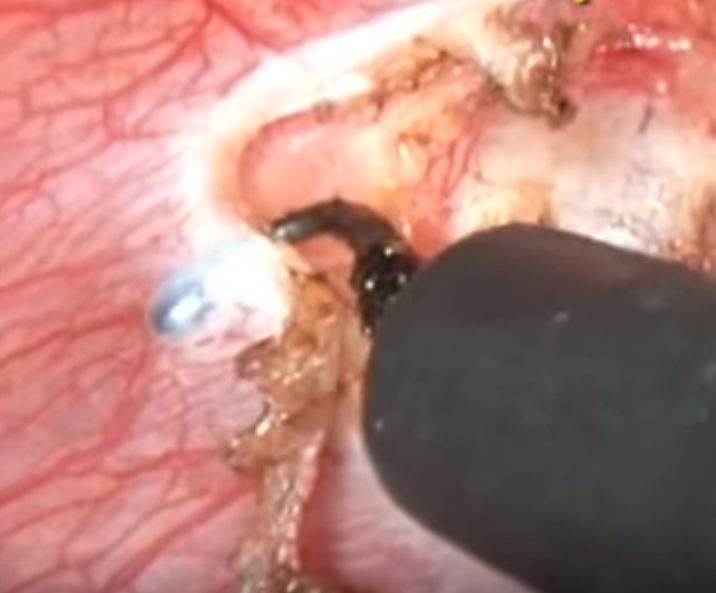
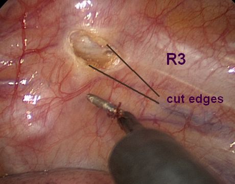
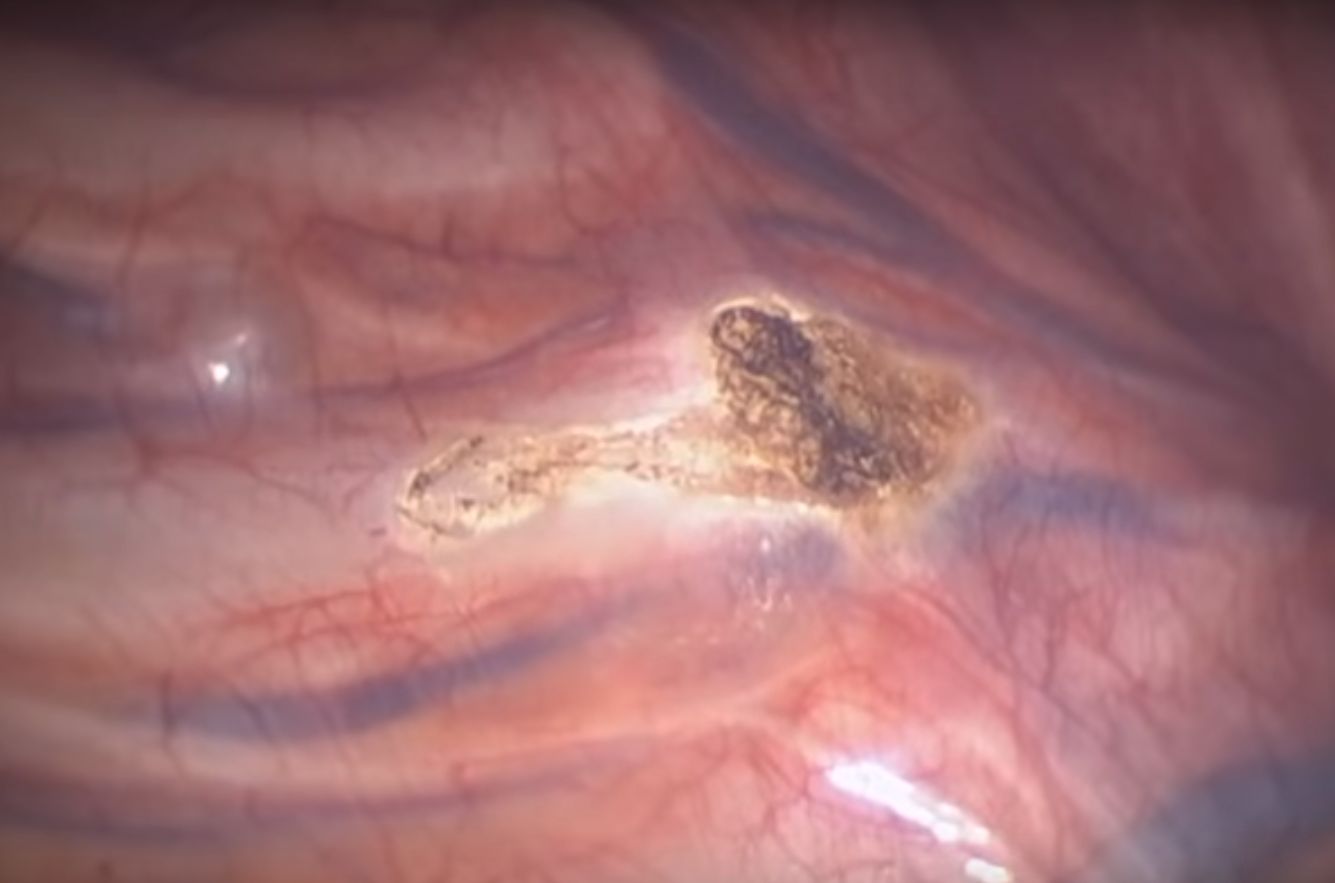
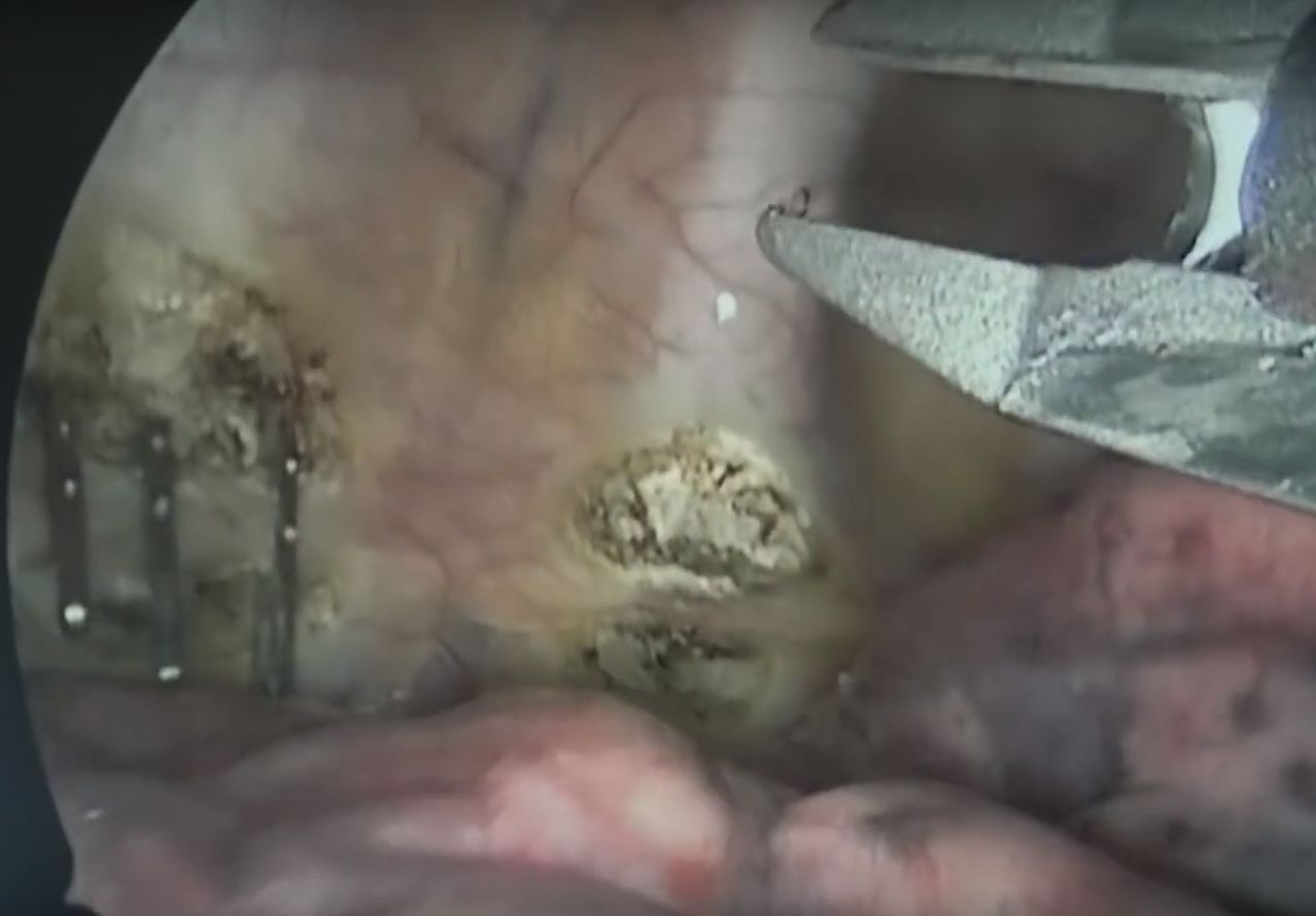
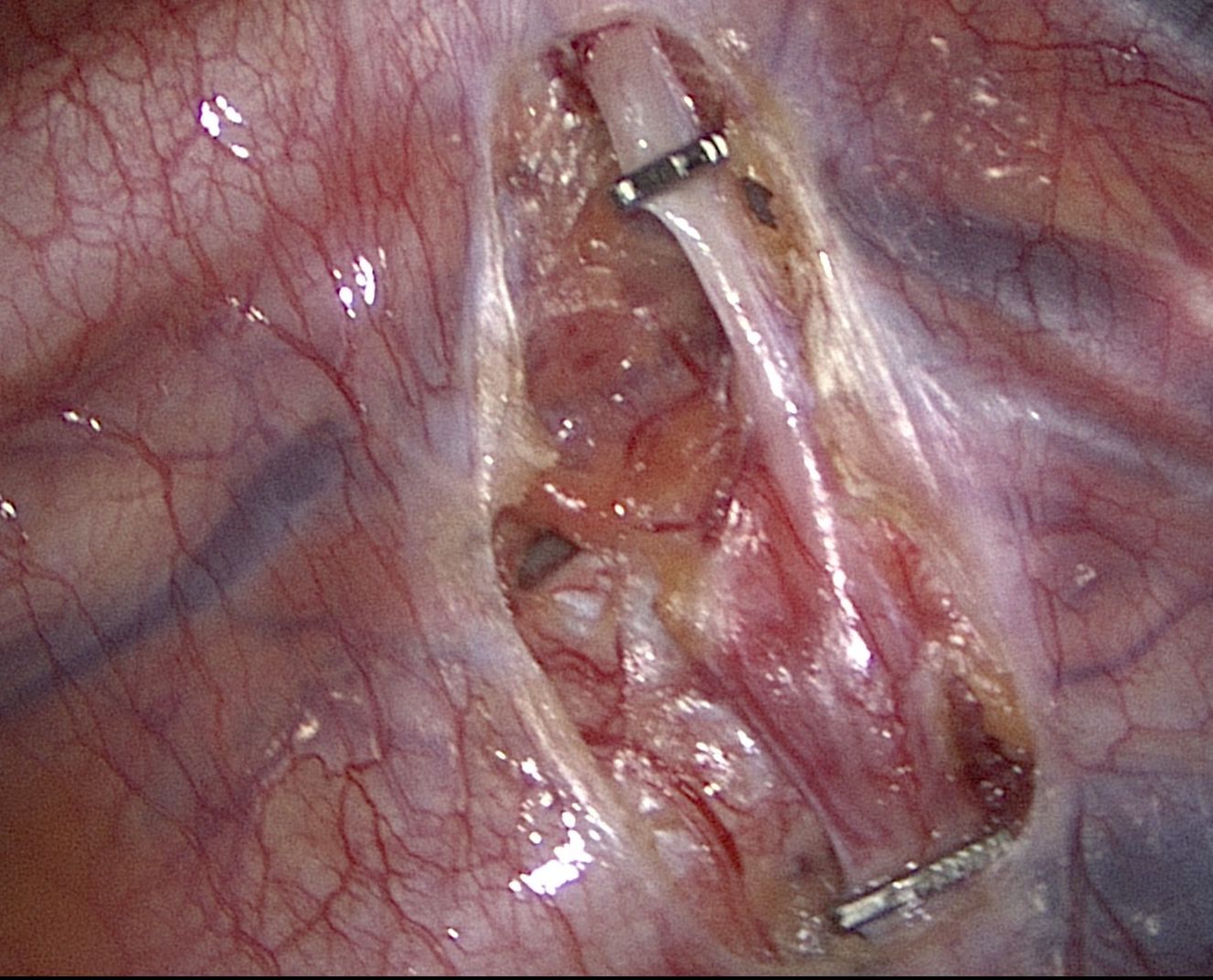
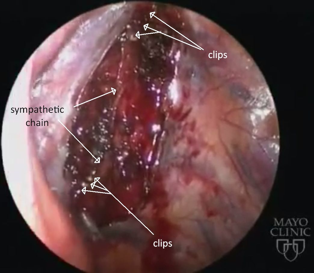
The ganglion is dissected free and exactly identified (in some cases, the ganglion can even extend beyond the lower edge of the 4th rib). Its upper end is always located below the lower edge of the 3rd rib (!). Interruption should be carried out at the interganglionic level, at the lower margin of the rib, not across any part of the ganglion. It is not clear, whether destroying the ganglion will add to the longterm efficacy.

Clips

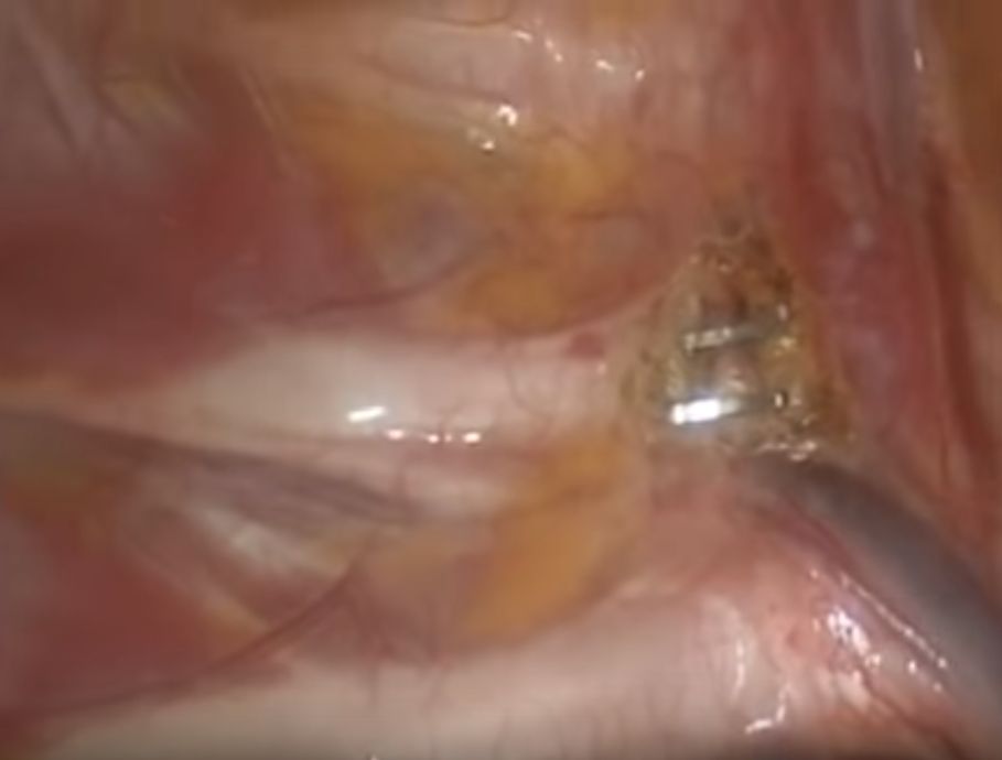
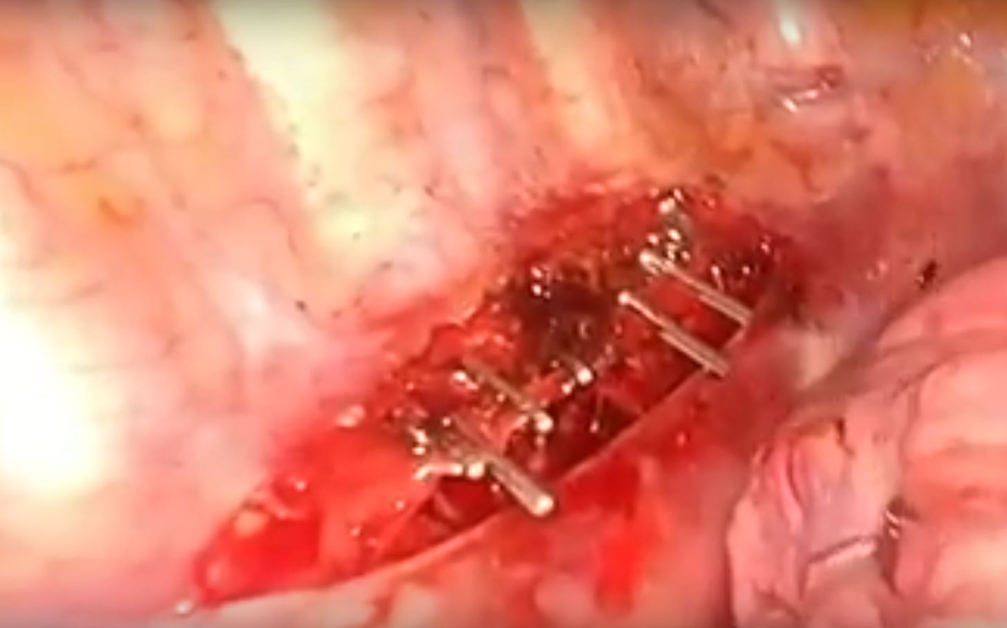
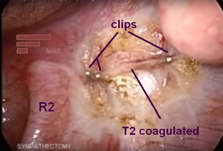
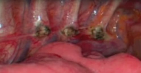
1 clip on lower margin of 3rd rib, 1 clip on lower margin of 4th rib (with or without additional clip on grey ramus communicans - grc) . The ganglion itself will remain intact and continue to be viable, but exiting connections will be blocked.

Clip above and below T3, grey ramus communicans (rcg) divided. Note location of ganglion covering 4th rib.


Analogous procedures with additional 3rd clip on grey ramus communicans (rcg).
Video

Electrocautery

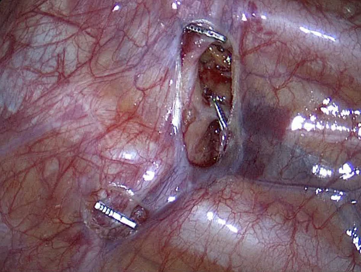
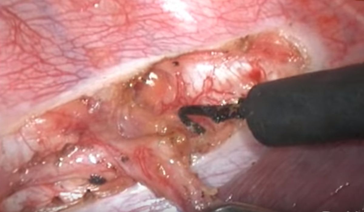
Ganglion is isolated by clean division (pure cutting current) at lower edge of 3rd rib and lower edge of 4th rib, careful (=soft) coagulation of the ganglion while lifting the ganglion from the underlying structures and keeping continuous awareness of the distribution of current density and heat development. This procedure is comparable to resection of the ganglion, but can be accomplished with a 2-port approach. Excellent method for palmar hyperhidrosis to achieve reliable long-term results with limited side-effects.

Note: upper and lower cut edge retracted after division, ganglion shrunk.
Video

Any other technique (use of blend current, division in other locations, fulguration, coagulation of ganglion while pressing against underlying structures, etc.) implies a significant risk of damaging adjacent ganglia and/or underlying structures (intercostal nerve, costovertebral joint, etc.).

Resection

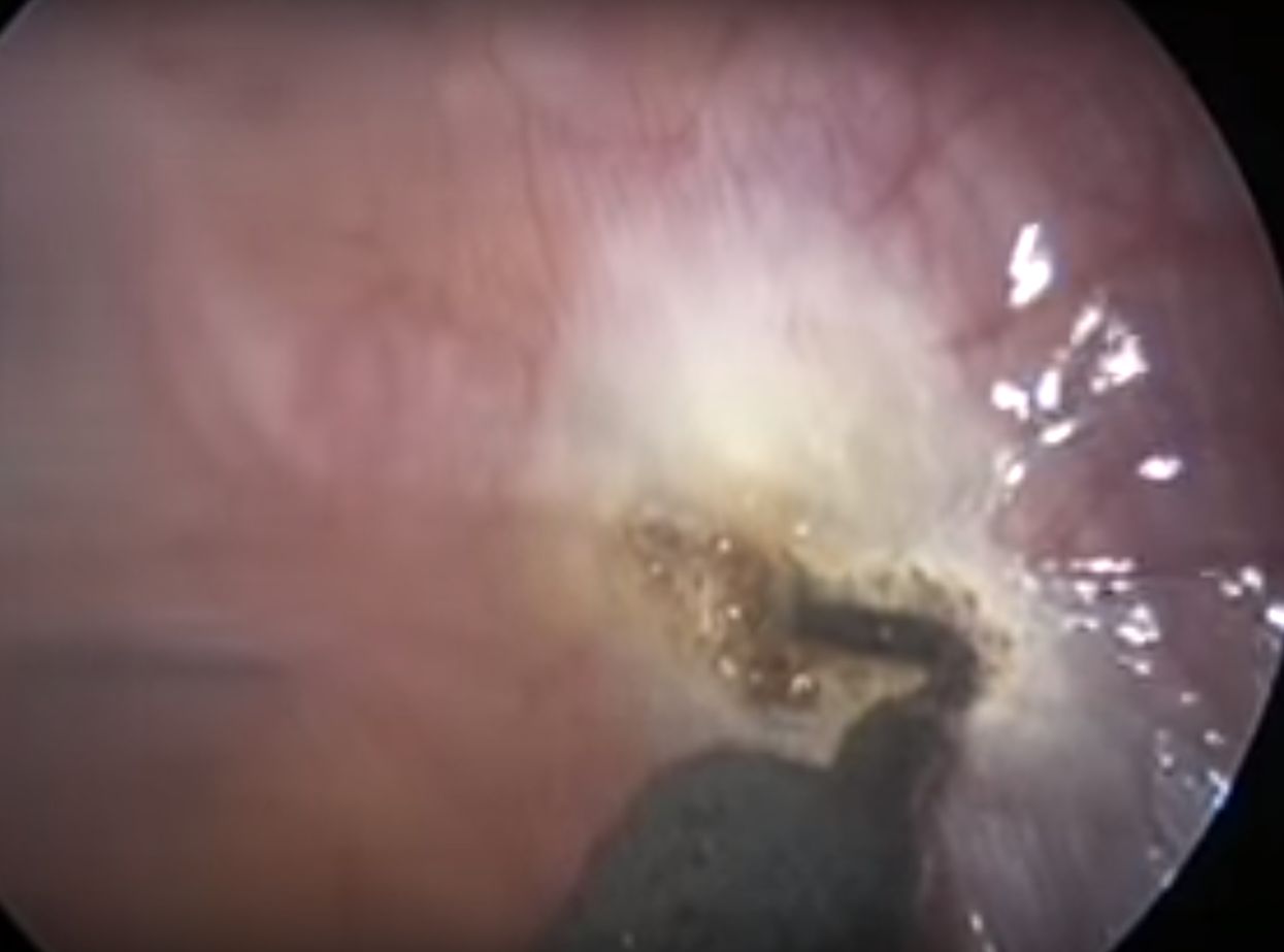
Clean division (plain electro-cut or scissors with application of clips to seal the cut-edge) at lower edge of 3rd rib and lower edge of 4th rib, excision of the ganglion, if necessary careful (soft) coagulation of bleeders without damaging adjacent or underlying structures.

T3 excised, from lower margin of 3rd rib to lower margin of 4th rib.

There are several variants to this technique (no seal, heavy coagulation due to bleeding, division in the middle or on top of the rib, etc) that carry always a risk for causing collateral damage and, consequently, post-operative discomfort and long-term side-effects.


T3-ganglion-resection using scissors only, without sealing or coagulating the cut edges. Risk for neuroma formation. The complete procedure is shown in the video.
Video

ETS T2-T3 (ganglion-oriented) ↰ 

This term implies blockade, destruction or removal of both ganglia T2 and T3.

As a rule, there is no rationale for eliminating both ganglia. It only risks adding or increasing the intensity of side-effects, without improving the desired outcome.

As with other variants of ETS, there are different surgical techniques in use to accomplish the task.

Severing the sympathetic chain above ganglion T2 and below ganglion T3 is followed by either coagulation or resection of the intervening segment containing the two ganglia.

Besides the superfluous destruction/excision of the T2 ganglion, collateral damage may occur if coagulation is used generously to stop bleeders or fulgurate the ganglia.

An odd variant of this principle consists in severing the rami communicantes of the ganglia T2 and T3, followed by blocking the chain above the T2 ganglion and at the level of the T3 ganglion, without resecting the segment.

Block with multiple clips above ganglion T2 and several clips on T3, after cutting all the rami communicantes. Given the anatomy of the T3 ganglion, part of it will end up crushed at random by the clips. The clinical result cannot be expected to be different from a resection of these ganglia. Again: very questionable the exclusion of the T2 ganglion, but presumably less harmful than leaving it partially functional.
Video

ETS T3-T4 (ganglion-oriented) ↰ 

Destruction or removal of both ganglia T3 and T4.

Sometimes used in patients with combined palmar and axillary hyperhidrosis. Generally, after a complete destruction/excision of the T3 ganglion, also sweating in the armpit ceases or is reduced to normal level. Including the T4 ganglion is considered by some surgeons to reduce the recurrence rate of axillary sweating.

As with other variants of ETS, there are different surgical techniques in use to accomplish the task.

Severing the sympathetic chain above ganglion T3 and below ganglion T4 is followed by either coagulation or resection of the intervening segment containing the two ganglia.

When resecting, collateral damage may occur if coagulation is used generously to stop bleeders or fulgurate the ganglia.

ETS T4 (ganglion-oriented) ↰ 

Neutralization of the T4 ganglion alone will, if carried out cleanly, regardless of technique (as described above -> ETS T3 ganglion-oriented), yields inconsistent results when used to treat palmar hyperhidrosis.

The author has no experience in treating isolated axillary hyperhidrosis with this method.

“Dirty” cuts on the 4th rib may damage the T3 ganglion sufficiently to “improve” the result, but such procedure has to be classified as rib-oriented R4, not as ganglion-oriented T4 procedure.

RESECTIONS  ↰ 

Resections imply removal of a section of the sympathetic chain.

Crucial elements of a resection are the following parameters:

  1. How the upper and lower division of the chain has been performed
  2. At what level the upper and lower cut has been performed
  3. How long segment has been removed
  4. By what means the communicating branches have been severed
  5. How bleeding vessels have been dealt with

Since the division and dissection of the nerve can be carried out by any of the above mentioned methods, the least traumatizing one should be chosen, keeping spread of thermal damage to a minimum. Anatomical boundaries of the chain should be respected, avoiding partial damage to a ganglion, including cutting through a ganglion.

Facial hyperhidrosis and facial blushing can be treated by one single interruption (cut or, preferably, clip). Ganglion resection is never indicated nor required.

Palmar or palmo-axillary hyperhidrosis is best dealt with by neutralizing the ganglion T3. This can be accomplished by resection or by careful coagulation of the ganglion, after detaching it at either end, and leaving it in place. Alternatively, the T3 ganglion can be efficiently isolated by applying titanium clips above, below and on the grey ramus communicans.

Resection of the T2 ganglion or extended excisions, involving multiple ganglia (like in the following examples), are almost never required and should be avoided.


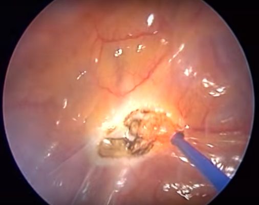
T2-T4 (cutting and blend mode electrocautery)

Resection of ganglia T2 and T4 (from rib 2 to 4th ICS) with cutting and blend current. Lower part of ganglion T4 left in place thermally injured. Useless carbonizing burns on ribs lateral to cut edge.
Video

T3-T5 (harmonic scalpel)

Resection of ganglia T3 to T5 with harmonic scalpel
Video

T2-T3 (blend mode electrocautery)

Resection of ganglia T2 and T3 with electrocautery (blend mode). Far too high energy setting, leading to uncontrolled heat propagation into and carbonizaton of surrounding tissues.
Video

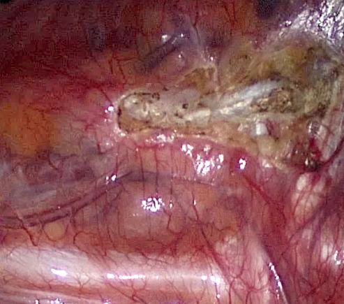
T2-T6 (left), T3-T7 (right) - Robot assisted sympathectomy (blend mode electrocautery): a questionable method.

The video displays a typical example of improper use of instruments and technology, excessive extent of destruction of sympathetic chain, systematic abuse of excessively high energy coagulating current with cutting properties, collateral damage to surrounding structures, apparent unawareness of the basic physical properties of electrocautery... In other words: a vivid example about how to make (a usually straight-forward minimal invasive) procedure overelaborate, messy and complicated, time-consuming, exaggerately traumatic and far too radical. And BTW: not symmetrical.
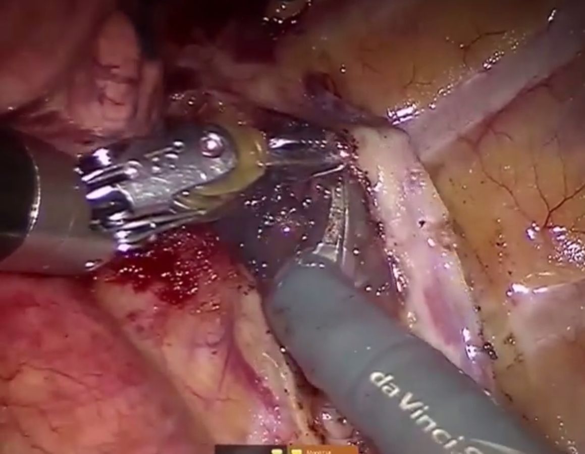
Right image: heat damage to the lung (lower left). The bad habit to use the grasping forceps for coagulation and, indeed, to cut(as shown in the video!), may result in unintentional heat injury to adjacent structures if the surgeon does not keep the unisolated surfaces of the instrument under control.
Video

Attention! When dividing the nerve with clean cutting current, broad tissue contact with electrode will disperse energy and result in soft coagulation instead of a cut:

Risky business! Over the 2nd rib, the chain is lifted with the hook electrode and cutting current is applied. Instead of the tip of the electrode, the whole hook surface is in contact with the nerve. The heat builds up too slowly in the tissue, tissue impedance remains low for too long and allows thermal energy to spread farther in craniad direction than intended, putting the stellate ganglion in jeopardy (note the whitish coagulated nerve above the electrode). Video


CONCLUDING REMARKS  ⇧ 

  • A ganglion is never located on the rib that carries the same number. It regularly starts a few millimeters below the lower edge of the rib. It often reaches the upper margin of the next-lower rib or even covers it, at least in part (e.g. ganglion T2 lies close to or stretches over the 3rd rib).
  • Clip application at the upper margin of the rib may partially squeeze the rostrally located ganglion, thereby risking to create a neurologically unstable focus.
  • A similar unstable nerve segment may result from applying coagulating (or blended) electrocautery current on the rib to divide the trunk.
  • Side-effects like bradycardia (low heart rate), hypotension (low blood pressure), unstable thermoregulation, extreme compensatory hyperhidrosis are mainly linked to partial damage or complete destruction of T2. Sympathectomy or block of ganglion T3, if performed with precision, without even touching the T2 ganglion, appears to reduce untollerable side-effects to almost nil, while palmar hyperhidrosis (and axillary) is reliably controlled. Unfortunately, many surgical procedures, defined as “ETS T3” or "R3" are interfering with the T2 ganglion, causing different degrees of mechanical or thermal damage to the ganglion, in most cases without even touching the T3 ganglion. In the majority of published peer-reviewed studies about sympathectomy, description of the procedure is far from precise or completely lacking. Needless to mention the confusion created when trying to interpret the results.
  • Interrupting the chain at the top of the 3rd rib, as recommended by the STS consensus, will put the T2 ganglion in jeopardy and should be avoided whenever possible to prevent severe side-effects. Therefore, any intervention on the nerve higher than the lower edge of the 3rd rib should be banned for hyperhidrosis of the upper limb.
  • Rib-oriented procedures will hit the real target (i.e. the sympathetic chain) in a fortuitous manner, since the anatomy of the chain itself is quite variable. In fact, the surgeon is disregarding the anatomical boundaries of ganglia and interganglionic segments. The wide array and heterogeneity of technical means and methods to carry out the interruption add to the uncertainty when trying to interpret the reports. In other words, with rib-oriented approaches, we can only vaguely guess what part of the sympathetic chain has been excluded and we have no indication to what extent the nerve has been damaged. Thence, the information from most reports is almost worthless, if the aim was to increase knowledge about the outcome of sympathetic surgery.
  • Even worse are those papers that give no closer description of the level and method employed, maybe mentioning just sloppily "T3" or "T2-4", or not even that - and there are plenty of such examples in the literature -, filling the journals with superficial information of little use.
  • There is no provocation in expressing these considerations, on the contrary, I consider it provocative that a large surgical community insists in treating a complex and delicate structure as the sympathetic chain blindly, without respect for its anatomy and its segmental boundaries, and using more often than not coarse methods to interrupt it, instead of treating it like a jewel. The sad conclusion is that the majority of publications on the subject is to be perused with considerable scepticism.
  • It is appalling to detect that, among publicly accessible videos, a majority exhibit excessively destructive methods and/or disregard for the anatomy of the sympathetic chain. Better not imagining that this may represent a projection for how ETS is performed worldwide. When scrutinizing these documents, one comes to the conclusion that there appears to be no difference between top teaching university hospitals and minor thoracic surgery units. Not even a large number of performed surgeries guarantees a better outcome for patients, if the method employed persistently replicates outdated or approximate technique. Unfortunately, most surgeons seem not aware of the basic physics of electrocautery and the depth of thermal damage caused far too high energy settings. Adding this harsh approach to the haphazard R2, R3 or R4 sympathicotomies (landmarks recommended by the STS-consensus), the surgeon has only a vague, if any, idea about what segment of the underlying sympathetic chain and to what extent it is damaged.
  • Among the multitude of methods dealing with interruption of the sympathetic chain, only a small fraction can be considered acceptable as being sufficiently accurate and delicate with the nerve, its ganglia and the surrounding structures. Most methods, widely used and many still in use, do not comply with the criteria of minimum traumatization, anatomically sound approach at correct level. As long as we do not recognize this, and as long every surgeon does it in "his" way, maybe performing only a couple of surgeries a year ("because it is that easy"), perpetuating methods described >20 years ago or using as tutorials some of the many videos ingenuously published on the net, we will continue to increase the number of patients with unfavorable outcomes or even destroyed lives.

ETS is an extremely valuable method to treat palmar hyperhidrosis, but it has to be done correctly, at the correct level, with precision, and using the least traumatic method available. The problem with ETS is not the principle itself, it is the way it is carried out. And unfortunately, with most of the methods described, the patient must rely on his/her luck not to end up with heavy undesired side-effects.

ESB (block with single clip) may also be a good solution for patients with debilitating facial ailments which do not respond to conventional therapy. However, the recurrence rate is higher for facial blushing, and, for facial hyperhidrosis, patient selection has to be particularly strict, because of the significantly higher risk for compensatory/reactive hyperhidrosis in this latter category of patients. Reversal (removal of the clip) will only lead to reduction of compensatory sweating if the nerve had not been completely crushed by the clip.


Technical hints and summary

  • The sympathetic chain is not a peripheral nerve with intersections that can be cut, clipped or coagulated with approximation. It should be considered as a series of miniature brain-like structures connected with short nerve segments. After mechanical or thermal trauma, the membrane of the nerve cells in these ganglia, conglomerations of nerve cells, may become permanently damaged and hyperpermeable. The ganglion may develop uncontrolled overactivity and an unstable behaviour, similar to an epileptic focus after brain injury. Different observations support the theory that this mechanism may contribute to or trigger severe undesirable side effects:
     a)  Patients, that have undergone reconstruction of the sympathetic chain with nerve graft, because of unbearable compensatory hyperhidrosis, report often improvement only days or weeks after surgery. The graft itself, however, would require several months to gain, if ever, any function. It seems reasonable to deduct that severing the nerve in healthy tissue, in order to bypass the scar and prepare a connecting point for the graft, seems to block exaggerated signal flow, originating from a pathologically active portion of the ganglion.
     b)  Another element pointing towards unstable ganglia is the surprising effect of membrane stabilizing anti-epileptic substances, found recently in selected patients with debilitating compensatory hyperhidrosis under off-label medication with topiramate.
     c)  Brock et al published a case in whom severe CS after "T2-3 ablation" was successfully treated with additional complete CT-guided percutaneous sympathicolysis of the T2 ganglion, which had presumably only been hurt at the previous sympathectomy.
    Brock M et al., Resolution of generalized compensatory hyperhidrosis related to sympathectomy after T2 sympatholysis. Eur J Cardiothorac Surg 2017(Dec); 52(6):1231-1232.
     d)  In the author's own experience, adopting an anatomically precise and technically accurate method has dramatically reduced the incidence of unbearable side effects in patients operated for palmar hyperhidrosis after 2001, compared to those who had undergone surgery in the decade before.
  • The interruption (cut or clip) should be executed with highest possible precision on the interganglionic segment, best in the middle of this segment in order to guarantee a safe distance to the adjacent ganglia.
  • A ganglion should be either preserved completely unharmed or, if needed, destroyed completely (after cutting its connections to the other ganglia), but never ever left partially damaged.
  • If the ganglia are not clearly visible through the pleura, the pleura should be incised along the entire length of the ganglion to determine its boundaries.
  • Use clips or cutting current, avoid coagulation (unless very carefully avoiding collateral thermal damage)
  • Interruption should strictly follow the anatomy of the sympathetic nervous system and not the underlying skeleton (ribs).
  • When treating palmar hyperhidrosis, ganglion T2 should be left untouched whenever possible.
  • For facial blushing and facial hyperhidrosis, one single clip on the interganglionic segment between T1 and T2 (lower edge of the 2nd rib) will do the job. Keep in mind the higher recurrence rate for FB, but additional surgical measures will not change the result. Consider also the higher indidence of troublesome CS in FHH (removal of clips required in 13% of cases).
  • Slight recurrence of moisture in the hands/feet is highly preferable to severe compensatory hyperhidrosis, therefore, in all cases, preference should be given to the most limited approach possible to reach a reliable result! If carried out correctly (clean neutralization of the T3 ganglion), the relapse rate is negligible (low single digit range).
  • When interrupting the sympathetic trunk “blindly” (i.e. using a rib-oriented approach, with or without opening the pleura over the trunk), severing the trunk at the lower edge of each rib will give best chance to keep clearance from the ganglia.
  • Avoid interruption over the rib (front surface) or at the top margin of the rib (the higher the interruption on the rib, the higher the probability of partial damage to the next higher ganglion).
  • Avoid neuroma formation (e.g. after cut with scissors without sealing the cut-edge).